
Gallbladder Ultrasound Scans Treatment For Gallstones (Naturally!)
an ultrasound exam to diagnose gallstones. Other imaging tests may also be used. • Ultrasound exam. Ultrasound uses a device, called a transducer, that. will be visible in the image. Ultrasound is the most accurate method to detect gallstones. • Computerized tomography (CT) scan. A CT scan is an x ray that produces

Focused Gallbladder Ultrasound YouTube
Gallstones, also called cholelithiasis, are concretions that may occur anywhere within the biliary system, most commonly within the gallbladder . Terminology Gallstones (cholelithiasis) describe stone formation at any point along the biliary tree. Specific names can be given to gallstones depending on their location:
Ultrasound River Radiology
top of page How are gallstones diagnosed and evaluated? Imaging is used to provide your doctor with valuable information about gallstones, such as location, size and effect on organ function. Some types of imaging that your doctor may order include: Abdominal ultrasound: Ultrasound produces pictures of the gallbladder and bile ducts.
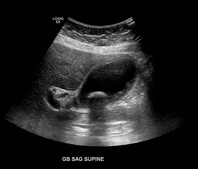
Gallstone ultrasound Radiology at St. Vincent's University Hospital
Treatment Preparing for your appointment Diagnosis Tests and procedures used to diagnose gallstones and complications of gallstones include: Abdominal ultrasound. This test is the one most commonly used to look for signs of gallstones. Abdominal ultrasound involves moving a device (transducer) back and forth across your stomach area.

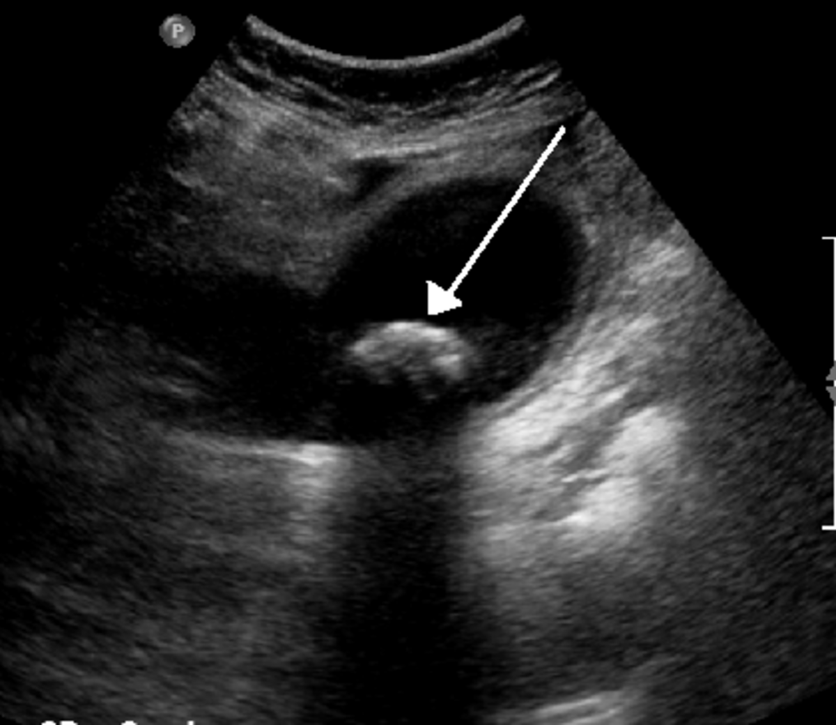
[Figure, Gallstone on PointofCare Ultrasound] StatPearls NCBI Bookshelf
Takeaway What is a gallbladder ultrasound? An ultrasound allows doctors to view images of the organs and soft tissues inside your body. Using sound waves, an ultrasound provides a real-time.
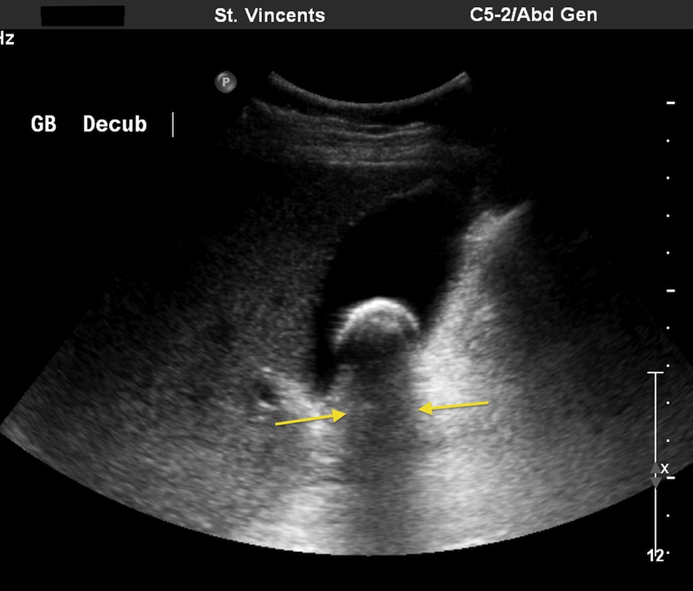
Multiple gallstones Radiology at St. Vincent's University Hospital
Normal findings on a gallbladder ultrasound include a thin-walled (<3 mm), anechoic, and pear-shaped structure that typically measures between 7-10 cm in length and 3-4 cm in width. The bile ducts should also appear anechoic without evidence of dilation or obstruction 7,8. Related pathology

Gallbladder Ultrasound Scans Treatment For Gallstones (Naturally!)
Products and services. This ultrasound shows gallstones in the gallbladder. Share. Tweet. Advertisement. Mayo Clinic does not endorse companies or products. Advertising revenue supports our not-for-profit mission. Advertising & Sponsorship. Policy.
Gallstone, Ultrasound Scan Photograph by Science Photo Library Pixels
1. Acute cholecystitis is one of the most common reasons for hospital admission with acute abdominal pain. 2. Approximately 90-95% of acute cholecystitis is related to gallstones, with 5-10% of cases due to acalculous disease. 3. Ultrasound is more useful than CT and MRI for the initial evaluation of acute biliary disease. 4.

Learn How to Spot Gallstones on Ultrasound YouTube
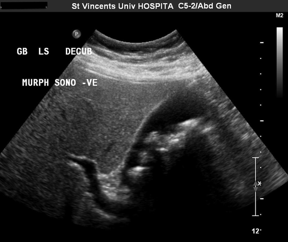
Introduction Symptomatic stones Asymptomatic stones Biliary colic Acute Hydrops Hydrops sign How to demonstrate an impacted stone Large diameter US-Murphy-sign US performed after the biliary colic Silent witnesses Reperfusion edema Acute Cholecystitis Differentiation Hydrops - Acute Cholecystitis CT in acute cholecystitis
Gallbladder Ultrasound Stones
Ultrasound uses sound waves to produce pictures of the gallbladder and the bile ducts. It is used to identify signs of inflammation involving the gallbladder and is very good at showing gallstones.. MRCP is a type of MRI exam that makes detailed images of the liver, gallbladder, bile ducts, pancreas and pancreatic duct. It is very good at.
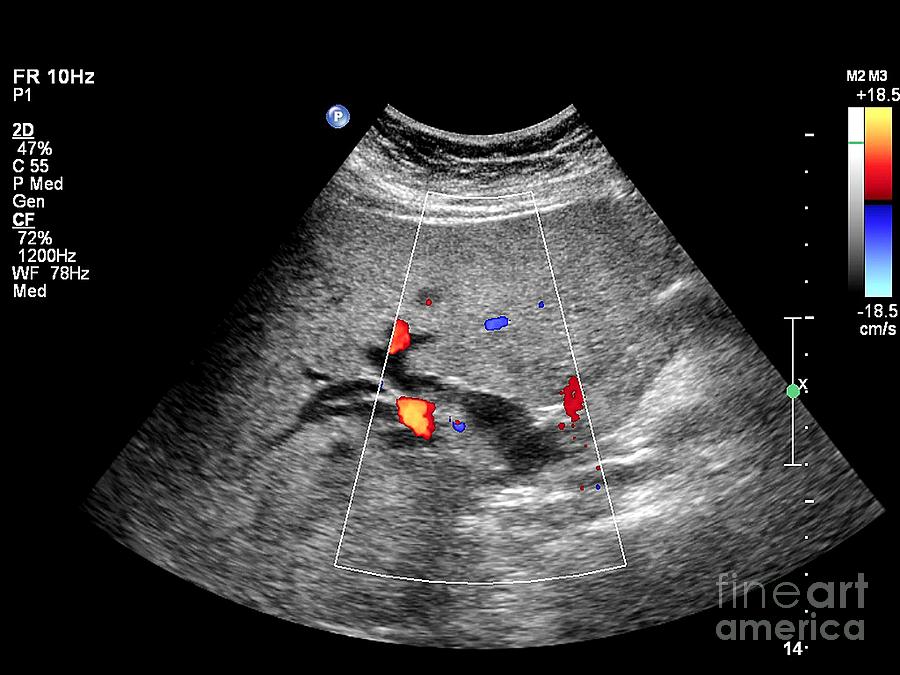
Gallstone, Ultrasound Scan Photograph by Science Photo Library Fine Art America
Ultrasound is the imaging modality of choice in patients with suspected gallbladder pathology. Indications include upper abdominal pain, right flank pain, jaundice and appropriate patients presenting with sepsis or septic shock.
Gallbladder Problems Gallstones HubPages
Gallstones are solid rounded particles composed of a combination of cholesterol and bilirubin that form within the gallbladder and within the biliary system. The size and number of gallstones is variable with some patients forming multiple small gallstones and others forming single or few large stones.
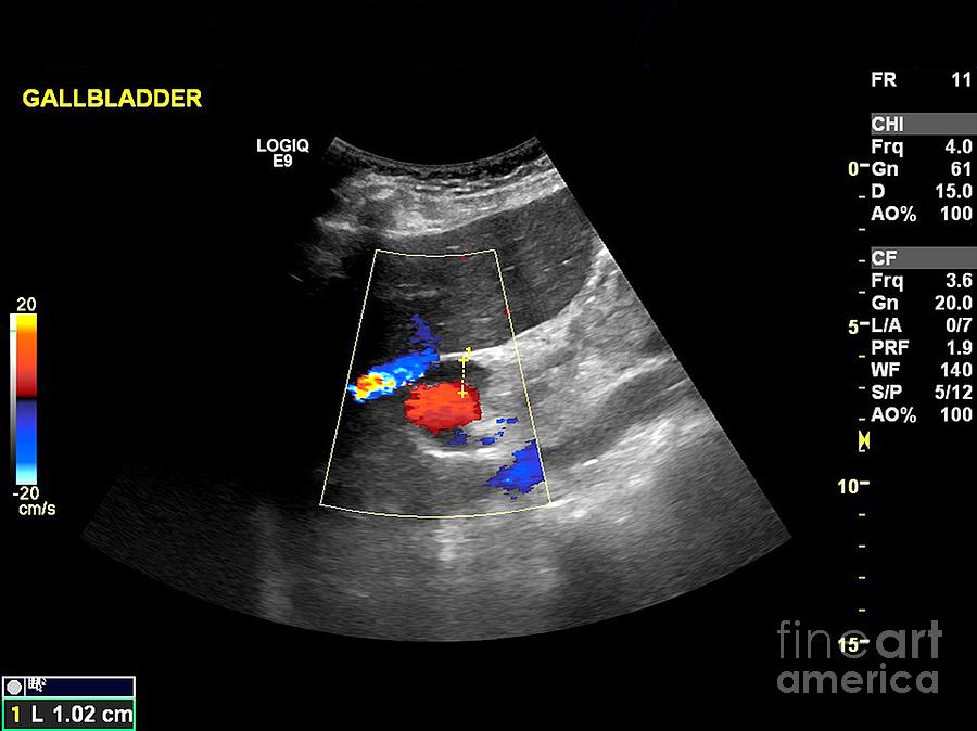
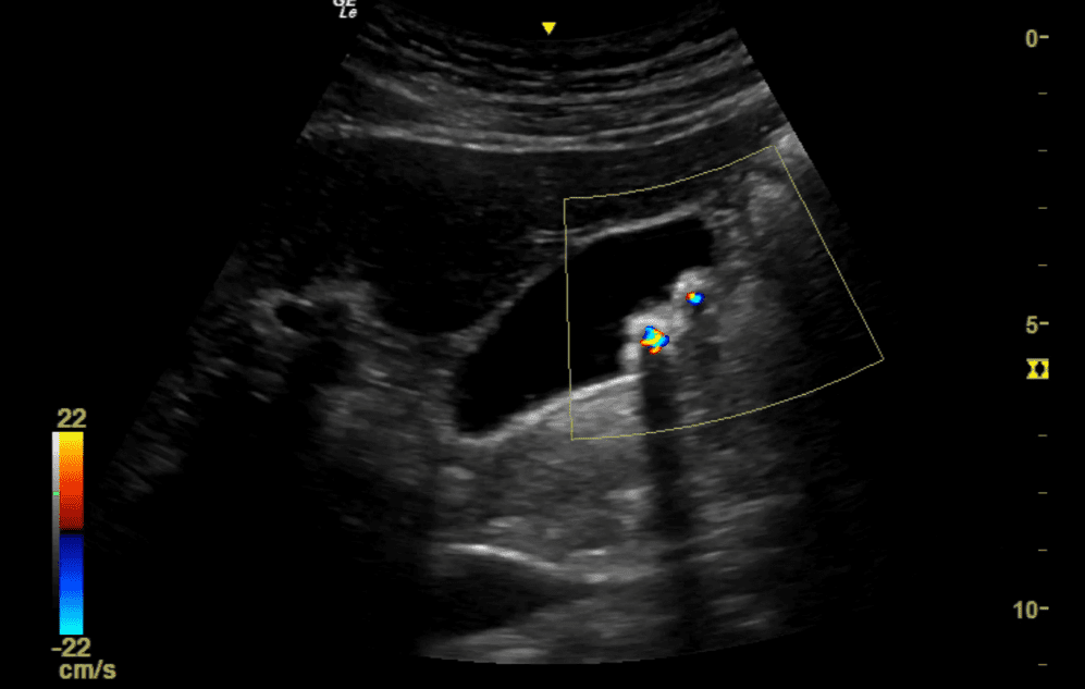
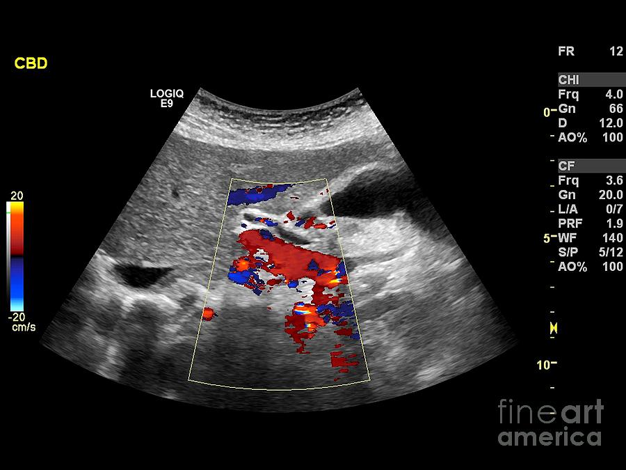
Gallstone, Doppler Ultrasound Scan Photograph by Science Photo Library Fine Art America
Imaging of the gallbladder for cholelithiasis and its complications has changed dramatically in recent decades along with expansion of interventional techniques related to the disease. Ultrasonography (US) is the method of choice for detection of gallstones. The characteristic US findings of gallstones are a highly reflective echo from the anterior surface of the gallstone, mobility of the.
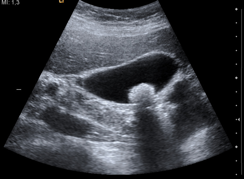
Gallstones, Ultrasound Scan Photograph by Science Photo Library
Ultrasound provides an optimal representation of the whole gallbladder (infundibulum, body, and bottom). A conventional transabdominal sonography image using low-frequency transducers shows a single or two layers of gallbladder wall (Fig. 2 a, b). High-resolution sonography (HRUS) using high-frequency transducers depicts three layers of gallbladder wall, including the innermost hyperechoic.

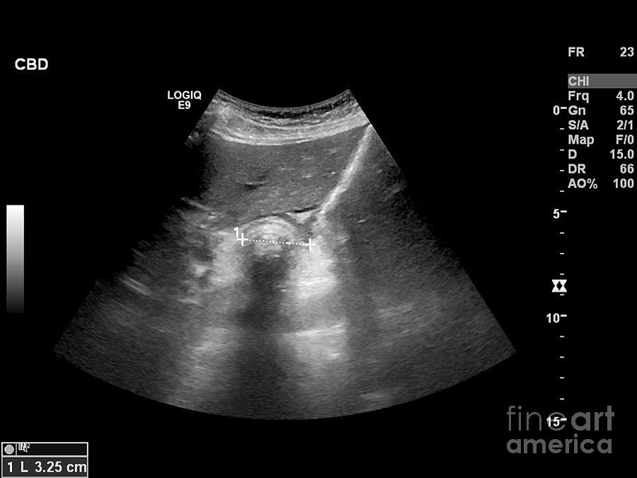
Gallstone, ultrasound scan Stock Image C018/7136 Science Photo Library
Imaging modalities Ultrasonography is the procedure of choice in suspected gallbladder or biliary disease; it is the most sensitive, specific, noninvasive, and inexpensive test for the.
Investigations GALLSTONES
Ultrasound is the best imaging test for finding gallstones. Ultrasound uses a device called a transducer, which bounces safe, painless sound waves off your organs to create an image or picture of their structure. If you have gallstones, they will be seen in the image. Sometimes, health care professionals find silent gallstones when you don't.
